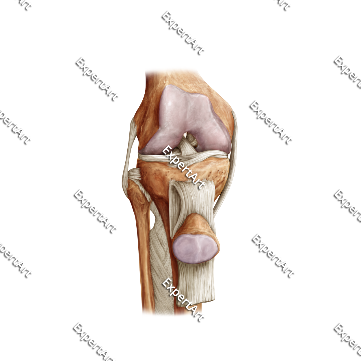
The knee is the joint in the body that binds the upper and lower leg, or the femur and tibia, respectively. Smaller bones, such as the tibia and the patella are also involved in the joint. The bones are joined together by ligaments that provide stability to the knee joint. These ligaments are the:
- Anterior cruciate ligament (ACL): this ligament prevents the tibia from sliding out under the femur.
- Posterior cruciate ligament (PCL): this ligament makes sure that the femur cannot slide forward too far over the tibia.
- The collateral ligaments: these two ligaments, the medial and lateral collateral ligament, prevent the femur from sliding to either side.
In addition to these ligaments, there are also tendons attached to the bones of the knee joint. These tendons connect the knee bones to the leg muscles and enable them to move the knee joint. These repetitive movements can have quite an impact on the knee, which is why there are two C-shaped pieces of cartilage that sit between the femur and tibia. These pieces of cartilage are called the medial and lateral menisci, and they function as shock absorbers between the tibia and femur. Besides the menisci, there are also numerous bursae present in the knee. These are fluid-filled sacs that reduce the friction between the moving parts of the knee joint.